UT Southwestern designs versatile 3D microscope for live cell imaging
The multi-immersion system switches between air, water and oil without rebuilding optical components.
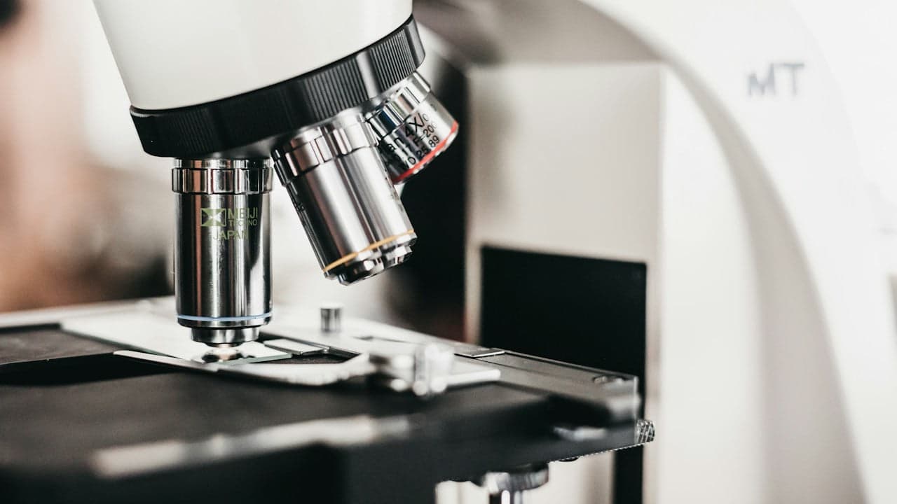
Researchers at UT Southwestern Medical Center have developed an invention that combines multiple light-sheet imaging modes into a single instrument. Reported in Nature Methods, the multi-immersion oblique plane microscope captures three-dimensional views of living cells and tissues while protecting them from light damage. The configuration allows research laboratories to run diverse biological experiments without purchasing separate specialized systems.
Switching between air, water and oil immersion
Standard light-sheet fluorescence microscopy typically requires dedicated hardware for specific experimental conditions. High-magnification views of intracellular details demand an oil-immersion objective lens. Observing thicker tissues or organoids requires water immersion or specialized immersion liquids. Scanning rapidly across culture plates without contacting the sample requires an air objective. According to research published by Phys.org, maintaining separate microscopes for each medium drives up capital costs and technical maintenance for research facilities.
To solve that constraint, corresponding author Reto Fiolka and co-first author Bingying Chen developed the multi-immersion oblique plane microscope, or miOPM. The new invention accommodates objective lenses tailored for air, water and oil on one optical chassis. An adjustable zoom lens compensates for refractive differences when scientists swap the fluid around a specimen, preserving image sharpness without requiring manual reconstruction of the internal optics.
“Our work makes a popular microscope technique, called light-sheet fluorescence microscopy, more versatile and user-friendly.”
— Reto Fiolka, Ph.D., associate professor at UT Southwestern Medical Center
Fast 3D imaging with limited light exposure
The instrument illuminates biological samples using thin sheets of light, scanning one plane at a time to build a three-dimensional model. This design restricts superfluous illumination, curbing phototoxicity and photobleaching so researchers can observe delicate living specimens over extended durations.
During operational trials described by Phys.org, the team imaged dynamic movements on immune cell surfaces and mapped organelle function. The system resolved individual mitochondria and mitochondrial DNA inside HeLa cervical cancer cells, actin networks inside pancreatic cancer cells, and carbohydrate-rich molecules within mouse lung tissue.
High-throughput screening on standard multiwell plates
Beyond isolated tissue preparations, the platform interfaces with multiwell formats, including standard 384-well plates. This capability allows automated scanning across hundreds of sample wells containing live human cells or mini-tumors. As the inventor team demonstrated, unifying these capabilities eliminates the need to recalibrate or switch between separate microscopes during high-throughput biological screens.
Pharmaceutical researchers can apply the innovation to evaluate how chemical libraries affect disease targets in 3D tissue models. Because the platform preserves cellular viability under repeated scans, researchers can follow disease-related structural changes in lung, liver and neurological models over time, highlighting how such intellectual property could benefit both academic laboratories and any biotech startup aiming to license the patent.
Read also on The Patent: Ancestral Enzyme Variant Generates ATP with Fewer Amino Acids · Engineers Solve Key Physical Hurdle in Exhaled Breath Diagnostics