GlioTrace uses artificial intelligence to track brain cancer cells in live tissue
Researchers at Uppsala University developed a computer vision method to map how glioblastoma cells switch movement styles across living brain slices
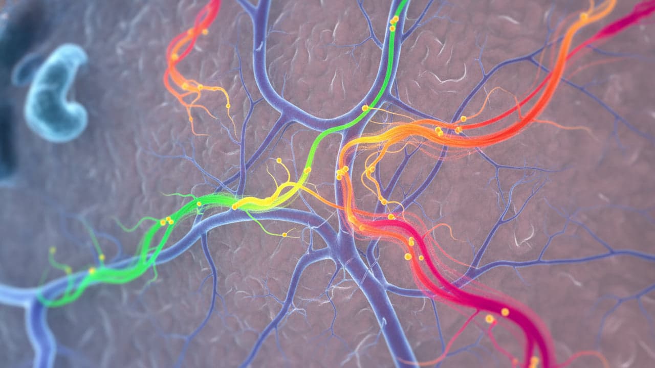
A computational imaging system called GlioTrace can track thousands of migrating glioblastoma cells across living brain slices for days at a time. Developed by scientists at Uppsala University, the method combines automated microscopy with artificial intelligence to reveal how individual brain tumor cells shift their shapes and change migration paths.
Automated microscopy captures live cell paths over five days
Glioblastoma remains one of the most aggressive forms of brain cancer, partly because tumor cells invade surrounding healthy tissue beyond the boundary of surgical removal. In an innovation aimed at studying this spread, researchers at Uppsala University used tumor cells taken from six patients and grew them in mouse brains. They cut the brains into thin slices and maintained them in laboratory cultures, according to findings published in Nature Communications and reported by MedicalXpress.
An automated microscope photographed the live tissue slices at intervals of no more than two hours for up to five days. Powered by artificial intelligence, GlioTrace tracked the individual cells across whole slices rather than small sample windows, recording the coordinates of each migrating cell from start to finish.
“What surprised us most was how flexible the tumor cells are. The same cell can change how it moves over just a few days. To develop treatments that stop tumor cells from spreading, we need to understand this ability to switch between different behaviors.”
— Madeleine Skeppås, Uppsala University
Machine learning identifies switching between migration states
The computational core of the invention was built by study co-lead author Madeleine Skeppås alongside researchers from Chalmers University of Technology and the University of Gothenburg. Their machine learning pipeline identifies individual cells, registers their shapes and tracks state transitions over time, as detailed in MedicalXpress.
The imaging revealed that glioblastoma invasion patterns differ between tumors. Some patient cells moved along blood vessels, while others migrated diffusely through brain parenchyma. Individual cells also proved capable of switching between independent migration and vascular crawling within days. Statistical models demonstrated that this behavioral switching helps cells navigate dense tissue environments.
Therapy testing reveals persistent fast-moving cell states
The research team tested how pharmaceutical compounds and standard treatments influence cell trajectories. In the experiments reported by MedicalXpress, two drug substances slowed down tumor cell invasion in the brain tissue slices. However, the exact response varied between patient samples and specific migrating cell types.
When exposed to combined radiation and chemotherapy, certain tumor models exhibited an increased fraction of fast-moving cells with a distinct shape. These cells occupied central nodes in the state transition networks, indicating they might persist during conventional therapy. Lead author Hitesh Mangukiya and professor Sven Nelander noted that the analysis code is open to other laboratories, though further testing is required before filing any patent or using the software to direct clinical decisions.
Read also on The Patent: GAO Warns USPTO Backlog Targets Risk Failure Under Staff Strain