Deep Tissue Microscopy Breaks the 100-Nanometer Barrier
Texas A&M researchers combine two optical methods to achieve 75-nanometer resolution inside thick biological specimens
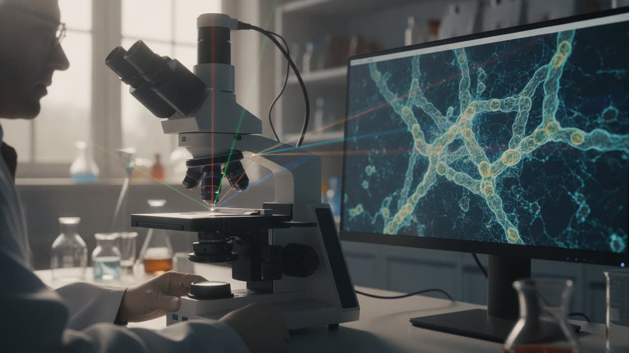
Biophotonics researchers at Texas A&M University have developed an optical imaging method that resolves structures smaller than 100 nanometers deep within intact biological specimens. The technique boosts standard two-photon microscopy resolution fivefold without sacrificing tissue penetration depth, as detailed in a study published in the Journal of Biomedical Optics.
Combining two methods to bypass the depth trade-off
Standard two-photon microscopy has long served as a staple tool for imaging living systems because it penetrates biological tissue with minimal cellular damage. However, looking deeper into samples has historically meant giving up fine detail. Surface-based super-resolution methods, such as stimulated emission depletion (STED) and photoactivated localization microscopy (PALM), can resolve structures well below 100 nanometers, but they fail to maintain that clarity when peering into thick tissues, according to a report by Phys.org.
The new optical invention bridges that performance gap. Developed by a team led by Aart Verhoef, assistant professor of biophotonics at Texas A&M University, the platform combines two optical protocols directly on a standard two-photon system: two-photon excited image scanning microscopy and super-resolution optical fluctuation imaging. The resulting hybrid system is designated as 2PE-ISM-SOFI.
“This technique builds on two microscopy techniques that were limited by depth and detail, but this technique allows us to look deep into tissue and improves the resolution.”
— Aart Verhoef, assistant professor of biophotonics at Texas A&M University
Achieving 75-nanometer resolution with quantum dots
By layering both techniques onto a single multiphoton setup, the research team achieved a lateral resolution of approximately 75 nanometers, roughly five times sharper than conventional two-photon systems. This scale corresponds to structures roughly a thousand times thinner than a human hair.
To demonstrate the prototype imaging setup, researchers analyzed cultured mouse ventral midbrain dopamine neurons. The team labeled cellular tubulin structures using QD625 quantum dots, which allowed optical fluctuation tracking under multiphoton excitation, as reported by Phys.org. Lead author Anton Classen and colleagues showed that the setup could resolve fine intracellular networks that typically blur into uniform masses under conventional deep-tissue multiphoton systems.
From neurology to plant biology
The breakthrough builds on preliminary experimental work that the Texas A&M team first published in 2024 in the journal PRX Life. While the latest study focused on neuronal structures, the underlying biophotonic innovation was designed to address practical observation limits across multiple scientific fields, including soil science and agriculture.
Co-author Alma Fernandez Gonzalez, a biophotonics researcher in the Texas A&M Department of Soil and Crop Sciences, noted that practical deployment across varied disciplines will require further technical refinement. Moving forward, the research team plans to adapt the optical system to investigate root networks, plant-microbe interactions and early disease pathology directly inside intact living plant tissues without the need for physical slicing, according to Phys.org.
Read also on The Patent: Mary Jo Ondrechen Receives the 2026 Ely S. Parker Award · Russia is conquering the space